The human foot is an engineering marvel, a complex structure designed to withstand immense pressure, facilitate movement, and provide essential support. However, conditions can arise that compromise its intricate mechanics, leading to pain, instability, and loss of function. One such critical issue is the collapse of the foot's arch, a common problem often addressed through specialized orthopedic interventions. Among these, FDL surgery, specifically a flexor digitorum longus tendon transfer, stands out as a highly effective solution for restoring vital arch support.
FDL surgery, which is short for Flexor Digitorum Longus surgery, is a pivotal procedure in foot and ankle orthopedics. While it has several applications, its primary role in addressing conditions that lead to the painful collapse of the foot's arch has cemented its importance. This procedure involves carefully repositioning or re-purposing the FDL tendon to bolster compromised structures, providing dynamic support where it's most needed. For more detailed information on its broader applications, you can explore FDL Surgery Explained: Foot & Ankle Tendon Repair.
Understanding the Flexor Digitorum Longus (FDL) Tendon
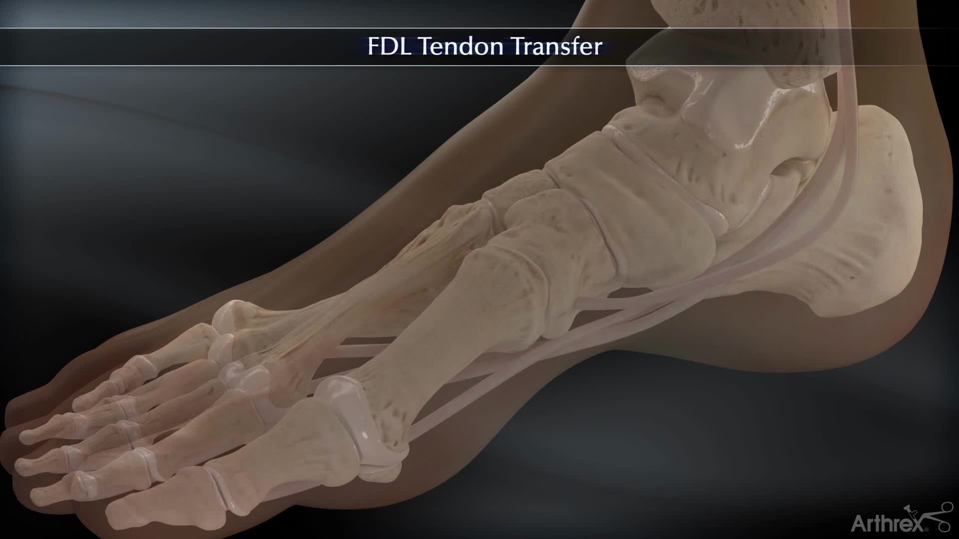
To appreciate the significance of FDL tendon transfer, it's crucial to understand the anatomy and function of this remarkable structure. The Flexor Digitorum Longus (FDL) is a deep muscle situated in the posterior compartment of the lower leg. From its muscular belly, a long, slender tendon extends down into the foot, embarking on a critical journey through intricate anatomical pathways.
Anatomy and Its Critical Role in Foot Function
As the FDL tendon descends into the foot, it wraps behind the prominent inner ankle bone, known as the medial malleolus. This strategic routing allows it to exert leverage crucial for foot mechanics. Upon entering the sole of the foot, the tendon elegantly branches into four separate slips. These slips then attach to the undersurface of the second through fifth toes, effectively connecting the muscle in the lower leg to the toes.
The primary function of the FDL muscle, facilitated by its tendon, is to flex the four smaller toes downward at their joints. This action is surprisingly important for stability, enabling us to grip the ground, maintain balance, and propel ourselves forward during walking or running. However, the FDL tendon's contributions extend beyond simple toe flexion; it plays a critical, albeit often underestimated, role in the overall structural integrity of the foot.
Beyond Toe Flexion: A Key Contributor to Arch Stabilization
In addition to its role in toe flexion, the FDL tendon significantly assists with plantarflexion – the motion of pointing the foot downward. More importantly, it acts as a dynamic stabilizer for the medial longitudinal arch, the primary arch along the inside of the foot. This arch is essential for shock absorption and maintaining the foot's biomechanical efficiency. When this arch falters, the entire foot structure can collapse, leading to significant pain and functional impairment.
A unique characteristic that makes the FDL tendon invaluable in reconstructive procedures is its "donor" potential. Due to the presence of other smaller muscles within the foot that also contribute to toe flexion, the FDL can be repurposed for a new role without causing a significant loss of toe movement. This biomechanical redundancy makes it an ideal candidate for tendon transfer surgeries, allowing surgeons to utilize a healthy tendon to replace a damaged or non-functional one, particularly in cases where the foot's arch support system has failed.
The Arch's Demise: Posterior Tibial Tendon Dysfunction (PTTD)
The most common and impactful reason for an FDL tendon transfer, especially when targeting arch support, is Posterior Tibial Tendon Dysfunction (PTTD). PTTD is recognized as the leading cause of adult-acquired flatfoot deformity, a progressive condition characterized by the gradual collapse of the foot's medial arch.
The posterior tibial tendon is the primary structure responsible for maintaining the foot's arch. When this critical tendon becomes inflamed, stretched, or torn, it loses its ability to support the arch effectively. Over time, this progressive weakening leads to the characteristic flattening of the foot, often accompanied by pain along the inner ankle, swelling, and a painful shift in foot mechanics. This condition doesn't just affect the arch; it can cause a ripple effect of deformities throughout the foot and ankle, making early intervention crucial.
In cases of PTTD, the FDL tendon transfer essentially acts as a replacement, stepping in to take over the vital function of the failed or severely weakened posterior tibial tendon. This procedure is a cornerstone in reconstructing the fallen arch and alleviating the associated symptoms. For more insights into how FDL surgery addresses various deformities, including flatfoot, you can read FDL Surgery: Correcting Flatfoot and Hammertoe Deformities.
The FDL Tendon Transfer Procedure for Arch Support
The FDL tendon transfer is a sophisticated surgical technique designed to create a new, dynamic support system for the collapsed arch. The procedure involves a careful rerouting and fixation of the healthy FDL tendon.
During the operation, the FDL tendon is harvested, detached from its original insertions, and then rerouted through a specific path. It is then securely fixed to the navicular bone, which is the historical attachment site of the posterior tibial tendon. This new anatomical position allows the transferred FDL tendon to exert a biomechanical force that actively pulls the arch upwards, effectively providing the much-needed dynamic support that was lost due to PTTD. The goal is not just to replace the tendon but to restore the active muscular pull that helps maintain the arch during weight-bearing and propulsion.
It's important to note that FDL tendon transfer for flatfoot correction is rarely performed in isolation. Adult-acquired flatfoot is a complex, multi-planar deformity that often requires a comprehensive surgical approach. Therefore, FDL tendon transfer is almost always performed in conjunction with other reconstructive operations. These concomitant procedures might include:
- Calcaneal Osteotomy: Where the heel bone (calcaneus) is carefully cut and repositioned to realign the foot and improve the overall weight-bearing axis.
- Lateral Column Lengthening: Lengthening the outside of the foot to correct a specific aspect of the flatfoot deformity.
- Gastrocnemius Recession: Lengthening the calf muscle to relieve excessive tension that can contribute to flatfoot.
- Fusion Procedures (Arthrodesis): In more severe or rigid cases, certain joints in the foot may be fused to provide permanent stability.
This multi-faceted approach ensures that all contributing factors to the flatfoot deformity are addressed, leading to a more stable, functional, and pain-free outcome for the patient.
What to Expect: Surgical Process and Recovery Insights
Undergoing FDL surgery for arch support is a significant step towards regaining foot health. Understanding the surgical process and the subsequent recovery phase is crucial for patients.
The Surgical Experience
FDL surgery is typically performed under general anesthesia, ensuring you are completely asleep and pain-free, or a regional nerve block, which numbs only the affected leg. The choice often depends on patient health, surgeon preference, and the complexity of the procedure. Depending on the extent of the surgery and the patient's individual needs, a hospital stay of one to two days may be required for initial monitoring and effective pain management immediately after the operation.
Post-Surgical Recovery and Rehabilitation
Recovery is a critical phase that significantly impacts the long-term success of FDL surgery. It demands patience, adherence to medical advice, and dedicated participation in rehabilitation.
- Immobilization: Immediately after surgery, your foot will be immobilized in a cast or surgical boot to protect the surgical site and allow the transferred tendon and any bone realignments to heal. This period is typically 6-8 weeks, during which weight-bearing will be restricted.
- Non-Weight-Bearing: You will likely be instructed to remain non-weight-bearing on the operated foot for several weeks. This means using crutches, a knee scooter, or a walker to get around. It's vital to strictly follow these instructions to prevent damage to the healing structures.
- Physical Therapy: Once partial weight-bearing is allowed, usually around 6-8 weeks, a structured physical therapy program will begin. This is paramount for recovery. Physical therapy will focus on:
- Restoring range of motion in the ankle and foot.
- Strengthening the muscles around the ankle and the newly transferred FDL tendon.
- Improving balance and proprioception.
- Gradual progression to full weight-bearing and functional activities.
- Pain Management and Swelling Control: Expect some pain and swelling post-surgery. Your medical team will provide medication to manage pain. Elevating your foot above heart level and applying ice (as directed) will help reduce swelling.
- Return to Activities: A gradual return to normal activities typically spans several months. Light walking may be possible by 3 months, with more strenuous activities like running or sports taking 6-12 months or even longer, depending on the individual and the extent of the surgery.
Practical Tip: Preparing your home environment before surgery can significantly ease your recovery. Remove tripping hazards, arrange essential items within easy reach, and consider setting up a comfortable recovery zone where you can elevate your foot. A strong support system from family and friends can also make a significant difference.
Conclusion
The Flexor Digitorum Longus tendon transfer is a testament to modern orthopedic innovation, offering a powerful solution for those suffering from debilitating arch collapse, primarily due to Posterior Tibial Tendon Dysfunction. By strategically repurposing a naturally redundant tendon, FDL surgery restores dynamic arch support, alleviates pain, and significantly improves foot function. While the journey through surgery and recovery demands commitment, the potential for a stable, pain-free foot and a return to an active lifestyle makes it a profoundly rewarding intervention. Consulting with an experienced foot and ankle orthopedic surgeon is the first step towards understanding if FDL tendon transfer is the right path for your specific condition.